Cardiovascular Suite is based on image processing of a B-mode ultrasound scans. The quality of the results can depend on the quality of the ultrasound image supplied to the system.
We recommend the use of a vascular probe with a frequency between 7MHz and 15MHz. The general settings of the ultrasound system must be those suggested by the manufacturer of the apparatus. It is important, however, exclude any noise reduction filters that could degrade the performance of the edge detection algorithm. In particular, it is important to exclude any time filters that cause a smoothing effect on the images in motion. These filters may have different designations (the most common name is persistence) depending on the model of ultrasound equipment. Please contact the manufacturer of ultrasound apparatus for information on how to exclude this type of filter.
 CAUTION: Exclude any noise reduction filter (especially temporal filters).
The artery should be viewed in longitudinal section and should be as horizontal as possible to the image. For Carotid Study we recommend an image depth of 3-4 cm.
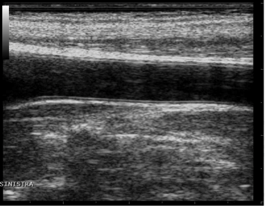
Example of carotid artery image
For FMD Studio we recommend an image depth of 2-3 cm. It is suggested also to choose a projection so that the vein is not visible (this normally appears immediately above the brachial artery). The algorithm for automatic tracking of the edges of the vessel could recognize the edge of the vein instead of the artery.
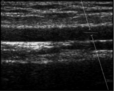
Example of brachial artery image