How to set up the ultrasound system
Cardiovascular Suite is based on image processing of a B-mode ultrasound scans. The quality of the results can depend on the quality of the ultrasound image supplied to the system.
The ultrasound device must have the following features:
The ultrasound device must be suitable for vascular imaging and it must be equipped with a vascular linear probe with frequency >= 5MHz (a range 7-15 MHz is recommended)..
The ultrasound device must have the B-mode imaging mode.
For the shear-rate measurement, the ultrasound device must have the Pulsed Wave Doppler (PWD) mode, and the B-mode and the PWD must be shown and updated simultaneously on the image (Dual mode).
For offline analysis, the ultrasound device must export in one of the following formats: AVI, MOV, MP4, DICOM, PNG, JPG, BMP, TIF. The size of the images must be <= 1920×1200 px.
For online analysis, the ultrasound device must have a video output in one of the following formats: S-Video or Composite Video in PAL or NTSC; VGA, DVI or HDMI with a resolution up to 1920×1200 pixels.
The B-mode in the image must have minimal resolution of 6 pixels/mm. If present, the size of the PWD window in the image must be at least 200x100 pixels.
The frame-rate of the video must be >= 17 frames per second.
The general settings of the ultrasound system must be those suggested by the manufacturer of the apparatus. It is important, however, exclude any noise reduction filters that could degrade the performance of the edge detection algorithm. In particular, it is important to exclude any time filters that cause a smoothing effect on the images in motion. These filters may have different designations (the most common name is persistence) depending on the model of ultrasound equipment. Please contact the manufacturer of ultrasound apparatus for information on how to exclude this type of filter.
CAUTION: The ultrasound device must be suitable for vascular imaging and it must be equipped with a vascular linear probe with frequency greater than 5MHz.
CAUTION: Exclude any noise reduction filter (especially temporal filters).
The artery should be viewed in longitudinal section and should be as horizontal as possible to the image. For Carotid Studio we recommend an image depth of 3-4 cm.
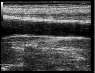
Example of carotid artery image
For FMD Studio we recommend an image depth of 2-3 cm. It is suggested also to choose a projection so that the vein is not visible (this normally appears immediately above the brachial artery). The algorithm for automatic tracking of the edges of the vessel could recognize the edge of the vein instead of the artery.
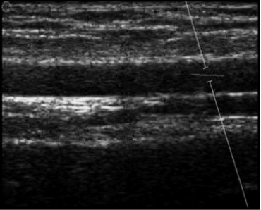
Example of brachial artery image
In addition, for FMD Studio, if you want to obtain both vessel diameter and shear rate, the ultrasound system must be in Duplex mode (simultaneous acquisition of B-mode and Doppler).
The angle between the Doppler beam and the vessel orientation should be ≤60 degrees. The sample volume should be as wide as possible but without encompassing the vessel walls and allowing for a slight margin for error in case of movement. Pay attention that the cursor of the doppler sample volume is not into the ROI where the diameter is computed. It is recommended that the sample volume is 5 - 15 mm apart from the ROI.
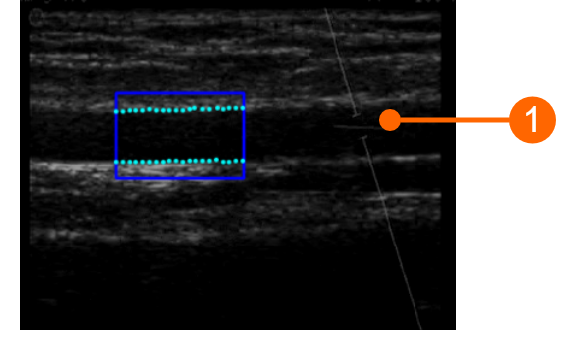
CAUTION: pay attention that nothing but the ultrasound image is into the ROI. Please note that the processing can be affected by annotations or any other graphical object that is superimposed to the image. In particular, pay attention that the cursor of the doppler sample volume is not into the ROI.
The scale of the Doppler flow profile should be set correctly on the ultrasound system. The vertical scale must be large enough to include the velocity profile during all the examination (in FMD measurements, greater velocity values are in reactive hyperemia). For the horizontal scale, we suggest a value of 3-4 seconds. Please note that the time average is computed over all the extend of the horizontal scale.
The Doppler Flow ROI must cover all the extent of the Doppler flow profile. The zero flow axis (2) must be included in the ROI: it will be automatically recognized and plotted in yellow. The vertical axis (3) must be external to the ROI. Please also ensure that any annotation (4) is outside the ROI since it could affect the flow analysis.
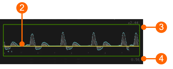
CAUTION: the processing can be affected by annotations or any other graphical object that is superimposed to the image into the Doppler Flow ROI.
Please remember that the tool for the calculation of the shear rate must be re-calibrated every time you change the size or scale of the Doppler flow profile. This calibration is present in FMD Studio analysis. It is recommended that the size or scale of the Doppler trace will be no longer changed once you have decided how to set up the ultrasound system.
FMD-Studio precision, expressed as coefficient of variation, is 10% for intra-observer intra-session measurements and 13% for intra-observer inter-session measurements of FMD%. For the Shear Rate measurement, the estimated precision is 2,3%.
Carotid Studio precision expressed as coefficient of variation is 2% for the diameter, 11% for the diameter variation during the cardiac cycle, 6% for IMT for intra-observer intra-session measurements and 3% for the diameter, 12% for the diameter variation during the cardiac cycle, 6% for IMT for intra-observer inter-session measurements. As regards geometric and statistics data the precision of the results expressed as coefficient of variation resulted lower than 10% for each measurement obtained on a single image by the same operator.